The Link Between CT Scans and Brain Cancer

By David W. Brown

CT (Computed Tomography) scans are tools that use ionizing X-rays to create cross-sectional images of the body. When directed at the head, they can reveal internal bleeding, tumors, and structural abnormalities in the brain. However, CT scans expose brain tissue to high levels of ionizing radiation, which can cause DNA damage in neural and glial cells.

Over time — and especially after repeated exposure — this radiation damage increases the risk of brain tumors, including gliomas and meningiomas. The association between CT scans and brain cancer has been confirmed by several large-scale epidemiological studies and mechanistic biological research.

A head CT scan typically delivers a dose of 1–2 millisieverts (mSv) to the body, but the absorbed dose to brain tissuecan be substantially higher, especially near the skull and scalp. Pediatric scans often deliver 50–60 milligrays (mGy)directly to the developing brain — an amount similar to what atomic bomb survivors experienced several miles from ground zero.

Children are especially vulnerable because:

  • Their brain cells are still dividing.
  • DNA repair mechanisms are less efficient.
  • They have longer lifespans, allowing latent cancers to manifest decades later.

Ionizing radiation from CT scans initiates several harmful biochemical and cellular processes that can culminate in brain tumor development:

a. DNA Double-Strand Breaks

High-energy X-rays eject electrons from atoms, breaking DNA strands.
If these breaks are misrepaired, they cause:

  • Mutations in critical genes (e.g., TP53, PTEN, EGFR).
  • Chromosomal rearrangements and genomic instability.
  • Activation of oncogenes or silencing of tumor suppressor genes.

b. Generation of Reactive Oxygen Species (ROS)

Ionizing radiation interacts with water molecules inside cells, forming ROS such as:

  • Superoxide anion (O₂•−)
  • Hydroxyl radical (•OH)
  • Hydrogen peroxide (H₂O₂)

These unstable molecules attack DNA, proteins, and lipids, compounding oxidative stress and furthering genetic instability — a known driver of tumorigenesis.

c. Epigenetic Changes

CT-induced radiation alters DNA methylation patterns and histone modification, changing gene expression without altering DNA sequences.
This can deactivate tumor suppressor genes or enhance oncogene activity, setting the stage for uncontrolled cell growth.

d. Bystander Effect

Even cells not directly struck by radiation become damaged through chemical signals (cytokines, nitric oxide) released from irradiated neighbors — expanding the affected area beyond the original beam path.

A. The Lancet Study – Pearce et al., 2012

  • Retrospective cohort of 178,604 British children who underwent CT scans between 1985–2002.
  • Researchers found:
    • Threefold increase in brain tumor risk among those who received cumulative brain doses of 50–60 mGy.
    • Proportional increase in leukemia risk at lower exposures (~30 mGy).
  • Crucially, these were diagnostic doses — not therapeutic radiation levels — proving that standard CT imaging alone carries measurable carcinogenic risk.

📘 Pearce, M. S. et al., “Radiation exposure from CT scans in childhood and subsequent risk of leukaemia and brain tumours,” The Lancet (2012).

B. The Australian Study – Mathews et al., 2013

  • Nationwide cohort of 680,000 young Australians exposed to CT scans before age 20.
  • Follow-up over 20 years revealed:
    • 24% higher overall cancer incidence in the CT group.
    • Significant increases in brain tumor rates (particularly gliomas and meningiomas).
    • Risk rose with the number of scans received.

📘 Mathews, J. D. et al., “Cancer risk in 680,000 people exposed to computed tomography scans in childhood or adolescence,” BMJ (2013).

C. Japanese Atomic Bomb Survivor Studies

  • Survivors who received comparable low-dose radiation exposures (10–100 mSv) showed similar latency and tumor patterns to those seen after medical imaging.
  • These findings support a linear no-threshold model, meaning any radiation dose carries some risk, even small ones from diagnostic scans.

📘 Preston, D. L. et al., “Solid cancer incidence in atomic bomb survivors: 1958–1998,” Radiation Research (2007).

  1. Gliomas – arising from glial cells, often linked to ionizing radiation.
  2. Meningiomas – benign or malignant tumors from the meninges; strong radiation sensitivity observed.
  3. Schwannomas – peripheral nerve sheath tumors occasionally reported after cranial imaging exposure.
  4. Pituitary adenomas – radiation can disrupt endocrine regulation, promoting pituitary cell proliferation.

Mechanistic Evidence:

  • Ionizing radiation induces mutations in NF2, TP53, and EGFR, all genes implicated in glioma and meningioma formation.
  • Radiation-induced gliomas often appear within 10–20 years after exposure, consistent with latency observed in survivors and CT-exposed children.

Latency and Dose–Response Relationship

Brain tumors caused by radiation typically appear after a latency period of 5–20 years.
The relationship between dose and risk is approximately linear — doubling the dose roughly doubles the risk.

A single head CT in childhood may increase absolute brain tumor risk by roughly:

  • 1 in 5,000 to 1 in 10,000 per scan (depending on dose and age).
    While this may seem small individually, at the population level (tens of millions of pediatric CTs per year), it translates to hundreds of preventable cancers annually.

Children’s Brains Are Especially Vulnerable

Children absorb higher doses because:

  • They are smaller, so the same X-ray energy penetrates more deeply.
  • Their tissues are more radiosensitive, as cells divide rapidly.
  • Neural stem cells in the developing brain are highly susceptible to mutations.

Additionally, cumulative exposure from follow-up scans for chronic conditions (e.g., epilepsy, trauma) multiplies long-term risk.

Adult Risks and Cumulative Exposure

While adults are less sensitive than children, repeated CT imaging over years still increases brain tumor risk.
Occupational studies in radiology staff and pilots exposed to chronic low-dose radiation have shown similar DNA damage markers, supporting the cumulative model.
Even “low-dose” protocols, when repeated, can surpass thresholds associated with carcinogenesis.

The Role of the p53 Tumor Suppressor Gene

Radiation-induced DNA damage often triggers activation of the TP53 gene, which governs DNA repair and apoptosis (“cellular suicide”).
If p53 function is overwhelmed or mutated, damaged cells survive and propagate, paving the way for tumor initiation.

This mechanism directly links CT-induced DNA damage to failure of the body’s natural cancer-prevention system — one reason the P53 gene is known as “the guardian of the genome.”

Safer Diagnostic Alternatives

To reduce risk, physicians should prioritize:

  • MRI scans (no radiation, ideal for soft tissue and brain imaging).
  • Ultrasound, when applicable for head and neck regions in infants.
  • Clinical observation and blood work before resorting to imaging.
  • Strict use of ALARA principles (“As Low As Reasonably Achievable”) for dose reduction.

The evidence linking CT scans to brain cancer is robust and biologically plausible.
Ionizing radiation from head CTs damages DNA, promotes oxidative stress, and triggers genetic and epigenetic changes that can lead to tumor formation years later.

Children and young adults face the greatest danger, but adults are not exempt — particularly those undergoing repeated or unnecessary scans.

Why CT Scans Are Harmful to the Body

By David W. Brown

Computed Tomography (CT) scans are among the most widely used diagnostic tools in modern medicine. They allow physicians to view the inside of the human body in cross-sectional images, providing crucial information that often cannot be obtained with ordinary X-rays or ultrasound. However, the benefits of CT imaging come at a significant cost: exposure to ionizing radiation. Unlike non-ionizing forms of imaging such as MRI or ultrasound, CT scans subject patients to doses of radiation strong enough to damage DNA, alter cellular processes, and increase the risk of long-term disease.

This article explores in depth the biological, biochemical, and systemic harms of CT scans, explaining the mechanisms of damage and why reliance on this technology should be approached with caution. While CT scans can be life-saving in emergency situations, their routine use presents avoidable risks to human health. See my previous articles on ionized radiation. I will also post an article on a detailed link between CT Scans and Brain Cancer coming next.

The Nature of CT Scans and Ionizing Radiation

CT scans use X-ray beams that rotate around the body, capturing multiple images that a computer processes into a detailed 3D view. The key issue lies in the fact that X-rays are ionizing radiation — electromagnetic waves with enough energy to knock electrons off atoms, creating charged ions.

When this ionization occurs in the human body, especially within living tissues, it disrupts the chemical bonds that hold DNA, proteins, and cellular membranes together. The consequences include:

  • DNA double-strand breaks, which are more difficult to repair than single-strand damage.
  • Mutations that can accumulate and trigger carcinogenesis.
  • Oxidative stress, caused by the generation of reactive oxygen species (ROS).
  • Cell death or malfunction, particularly in sensitive tissues such as bone marrow or reproductive organs.

Thus, each CT scan carries a biological cost, even if the harm is not immediately visible.

Radiation Dose: How Much Is Too Much?

The severity of harm from CT scans depends largely on the radiation dose delivered. Medical researchers measure radiation exposure in units called millisieverts (mSv). For comparison:

  • A standard chest X-ray exposes a patient to about 0.1 mSv.
  • A head CT scan can deliver 2 mSv.
  • An abdominal CT scan may expose patients to 8–10 mSv.
  • Some complex scans (e.g., cardiac CT angiography) can exceed 15–20 mSv.

To put this in context, the average person is naturally exposed to 3 mSv per year from background radiation in the environment. A single abdominal CT, therefore, can equal three years of natural exposure delivered in seconds.

Repeated scans amplify the risk dramatically. Patients with chronic conditions who undergo multiple CT scans may accumulate exposures equivalent to hundreds of chest X-rays, placing them in a significantly higher risk category for radiation-induced disease.

DNA Damage and Cancer Risk

The most concerning harm from CT scans is their potential to induce cancer. Ionizing radiation is a well-established carcinogen, classified by the World Health Organization’s International Agency for Research on Cancer (IARC) as a Group 1 carcinogen.

Pathways of Cancer Induction:

  1. DNA Double-Strand Breaks (DSBs): When high-energy X-rays strike DNA, they can sever both strands of the helix. Improperly repaired DSBs result in mutations, deletions, or chromosomal translocations.
  2. Epigenetic Alterations: Radiation can silence tumor suppressor genes (like p53) or activate oncogenes through methylation changes.
  3. Oxidative Stress Cascade: Radiation stimulates the production of free radicals, which oxidize DNA bases and further destabilize genetic integrity.
  4. Bystander Effect: Even non-irradiated neighboring cells can become cancer-prone through chemical signals sent by irradiated cells.

Large population studies confirm this danger. For instance, research on children exposed to CT scans has shown increased risks of brain tumors and leukemia, with the risk correlating with the cumulative dose. Children are especially vulnerable because their cells divide more rapidly and their lifespan allows more time for radiation-induced cancers to develop.

Effects on Specific Organs and Systems

Brain

CT scans of the head are common in cases of trauma, but ionizing radiation is particularly harmful to brain tissue. Research indicates radiation can alter neuronal stem cell populations, contributing not only to cancer but also to subtle cognitive impairments over time. See my next detailed article on the link between Brain Cancer and CT Scans. 

Thyroid

The thyroid gland, located near the surface of the body, is highly sensitive to radiation. Even relatively low doses can increase risks of thyroid cancer, especially in children and adolescents.

Lungs

Chest CT scans expose lung tissue to high doses. This is concerning because lung tissue is highly vascularized and prone to DNA damage accumulation. Lung cancer risk rises proportionally with repeated exposure.

Reproductive System

The ovaries and testes are extremely sensitive to radiation, and exposure can impair fertility. Germ cell mutations may even affect future generations.

Bone Marrow

As the cradle of immune and blood cell production, bone marrow is especially vulnerable. CT radiation can damage stem cells, raising risks for leukemia and other blood disorders.

Non-Cancer Health Effects

Beyond cancer, CT scans contribute to a range of other health issues:

  • Cataracts: Radiation exposure to the eyes can cloud the lens, leading to premature cataract formation.
  • Cardiovascular Damage: Radiation-induced inflammation in blood vessels contributes to atherosclerosis and heart disease.
  • Immune Suppression: Damage to white blood cells and bone marrow may reduce immune system function.
  • Accelerated Aging: Cellular senescence, triggered by DNA damage, leads to premature aging of tissues.

Vulnerable Populations

Some groups face disproportionate harm from CT scans:

  1. Children: Rapid cell division and longer expected lifespan make children more sensitive to radiation.
  2. Pregnant Women: CT radiation can harm fetal development, increasing the risk of birth defects or childhood cancers.
  3. Patients with Chronic Illnesses: Those who undergo multiple scans over time accumulate high doses.
  4. Healthcare Workers: While shielded, workers around CT equipment may experience low-level occupational exposure.

The Illusion of Safety in “Low-Dose” CT

In recent years, manufacturers have introduced so-called “low-dose CT” technology, especially for lung cancer screening. While doses are lower than traditional CT, they are still significantly higher than ordinary X-rays. Moreover, repeated annual screening adds up, nullifying the “low dose” advantage. There is no truly safe level of ionizing radiation — even the smallest dose increases cancer risk according to the linear no-threshold model.

Alternatives to CT Scans

Safer imaging options exist for many situations:

  • Ultrasound: Uses sound waves, with no radiation. Effective for soft tissue and fetal imaging.
  • MRI (Magnetic Resonance Imaging): Uses magnetic fields and radio waves, providing detailed images without radiation.
  • Physical Examination and Blood Testing: Sometimes overlooked, but can reduce reliance on imaging altogether.

Unfortunately, CT scans are often chosen for convenience, speed, and availability rather than true medical necessity.

Overuse and Industry Influence

Another harmful aspect of CT scans lies in their overuse. Studies show that up to 30% of CT scans may be medically unnecessary. Factors driving this overuse include:

  • Defensive medicine: doctors ordering tests to avoid liability.
  • Financial incentives: hospitals profit from expensive imaging procedures.
  • Patient demand: people equating more imaging with better care.

This systemic overuse multiplies the radiation burden on the population, raising public health risks unnecessarily.

Long-Term Public Health Implications

The widespread use of CT scans contributes to a silent but significant public health burden. Some estimates suggest that 1–2% of all cancers in developed countries may be linked to CT scan exposure. Given the billions of scans performed worldwide, the cumulative radiation exposure to the global population is staggering.

If safer diagnostic alternatives were prioritized, this preventable burden could be reduced dramatically. Instead, CT scans remain routine, embedding long-term cancer risk into standard medical practice.

By exposing patients to ionizing radiation, they cause DNA damage, oxidative stress, immune disruption, and increase the risk of cancers and other diseases. Vulnerable populations such as children, pregnant women, and chronically ill patients bear the greatest risk. The illusion of safety from “low-dose” CT scans and the systemic overuse of this technology further amplify the harms.

Patients and physicians alike should critically evaluate whether a CT scan is truly necessary or whether safer alternatives can provide the needed information. Only by reducing unnecessary exposure can society limit the hidden epidemic of radiation-induced disease tied to medical imaging.

Controlled Ionization in the Human Body

By David W. Brown 

Ionization inside the body refers to the generation and regulation of charged particles (ions) during normal biological processes. Unlike harmful uncontrolled ionization from radiation exposure, controlled ionization is a fundamental and safe part of life. Every cell relies on carefully regulated ion flows to maintain function, communicate, and generate energy.

1. Ion Channels and Membrane Potentials

  • Cell membranes contain protein structures called ion channels that allow selective passage of sodium (Na⁺), potassium (K⁺), calcium (Ca²⁺), and chloride (Cl⁻).
  • By moving ions in and out, cells create an electrical gradient (the membrane potential).
  • This “controlled ionization” is essential for:
    • Nerve impulses (action potentials) – rapid Na⁺ inflow followed by K⁺ outflow
    • Muscle contraction – triggered by Ca²⁺ release from storage vesicles
    • Hormone release – ion flows stimulate vesicle fusion and secretion

This is tightly regulated; uncontrolled ion leakage disrupts signaling and can lead to cell death.

2. Ionization in Energy Metabolism

  • The mitochondria (the cell’s power plants) generate ATP through oxidative phosphorylation.
  • As electrons move along the electron transport chain, protons (H⁺ ions) are pumped across the inner mitochondrial membrane.
  • This creates a proton gradient (a controlled ionization state), which drives ATP synthase to convert ADP into ATP.
  • This is one of the most important controlled ionization events in biology — turning food into usable cellular energy.

3. Calcium Ionization as a Messenger

  • Calcium (Ca²⁺) is a “universal signal ion.”
  • Inside cells, Ca²⁺ levels are normally kept extremely low.
  • Controlled release of Ca²⁺ from storage (endoplasmic reticulum) triggers:
    • Muscle fiber contraction
    • Neurotransmitter release in the brain
    • Fertilization (egg activation by sperm entry)
    • Blood clotting cascades

If ionization is uncontrolled, calcium floods the cell and causes damage (excitotoxicity, necrosis).

4. Controlled Ionization in Blood and Fluids

  • Blood plasma contains precisely regulated ion concentrations:
    • Sodium: ~135–145 mmol/L
    • Potassium: ~3.5–5 mmol/L
    • Calcium: ~2.2–2.6 mmol/L
  • Kidneys and hormones (like aldosterone and parathyroid hormone) tightly control these levels.
  • This prevents arrhythmias, seizures, and muscle dysfunction.
  • Even tiny imbalances in ionized calcium or potassium can be life-threatening.

5. Redox Ionization in Antioxidant Defense

  • Many molecules undergo controlled ionization-reduction cycles:
    • Glutathione (GSH ↔ GSSG) acts as a redox buffer
    • Vitamin C (ascorbate ↔ dehydroascorbate) cycles between ionized states
  • These processes neutralize free radicals while preventing runaway oxidative stress.
  • The body essentially “uses ionization” as a shield against uncontrolled molecular damage.

6. Controlled Ionization in pH Balance

  • The human body regulates hydrogen ion (H⁺) concentration to keep blood pH at ~7.35–7.45.
  • Buffers (bicarbonate system), lungs (CO₂ exhalation), and kidneys (H⁺ excretion) maintain this.
  • Too much H⁺ = acidosis; too little = alkalosis.
    Both disrupt enzyme function, proving how critical controlled ionization is.

7. Immune System and Ionization

  • Immune cells (like neutrophils) use a controlled burst of ionized radicals (reactive oxygen species, ROS) to kill pathogens.
  • This oxidative burst is highly targeted — designed to kill microbes without harming host tissue.
  • Antioxidants inside the cell prevent “spillover” damage.

In the human body, controlled ionization is not only natural but vital. It includes:

  1. Ion channel regulation in nerves, muscles, and glands
  2. Proton gradients in mitochondria for ATP production
  3. Calcium ionization as a signaling messenger
  4. Plasma ion balance maintained by kidneys and hormones
  5. Redox ionization in antioxidant defense
  6. Hydrogen ion control for pH balance
  7. Immune cell ionization bursts for pathogen destruction

When kept under tight control, ionization sustains life. When uncontrolled (e.g., radiation damage, electrolyte imbalance), it threatens health.

Ionization and Its Harmful Effects on the Human Body

By David W. Brown

Ionization is the process where atoms or molecules gain or lose electrons, creating charged species known as ions. In the human body, controlled ionization is vital for normal function—nerve impulses depend on sodium and potassium ions, and energy metabolism relies on electron transfers. But when ionization is uncontrolled, it can damage DNA, proteins, lipids, and cells, driving disease, premature aging, and cancer.

A major source of confusion among the public is the difference between ionizing radiation and non-ionizing radiation. Ionizing radiation—such as X-rays, gamma rays, and radioactive particles—has enough energy to strip electrons from atoms, breaking chemical bonds. This is dangerous to living tissue. Microwave ovens, on the other hand, use non-ionizing radiation. Microwaves cause molecules—primarily water—to vibrate, generating heat, but they do not have enough energy to ionize atoms or damage DNA directly. Understanding this distinction is critical when talking about ionization and its harmful effects. In my book Taste Versus Cancer, I explore Reactive Oxygen Species (ROS) and hydroxyl radicals in greater depth.

What Is Ionization?

The Basics

Ionization occurs when an atom or molecule loses or gains an electron:

  • Cations are positively charged (lost electrons).
  • Anions are negatively charged (gained electrons).

In biology, controlled ionization is normal and beneficial:

  • Nerve conduction relies on waves of Na⁺, K⁺, and Ca²⁺ moving in and out of neurons.
  • Energy production in mitochondria depends on electron transport chains where ionization and reduction happen step by step.
  • Detoxification in the liver often requires ionization of toxins to make them more water-soluble There is right there I think so he’s fast you can do everything with them for elimination.

Trouble starts when ionization happens randomly and excessively, often due to high-energy exposures like ionizing radiation.

Sources of Harmful Ionization

Ionizing Radiation

This is the main culprit behind harmful ionization in biology. Examples include:

  • Medical: X-rays, CT scans, radiotherapy.
  • Environmental: Radon gas, cosmic rays, radioactive fallout.
  • Occupational: Nuclear workers, radiology staff, frequent airline crews.

Ionizing radiation carries enough energy to knock electrons loose from DNA, proteins, and lipids, causing chain reactions of damage.

Chemical and Environmental Sources

Even without nuclear radiation, some exposures can cause excessive ionization:

  • Heavy metals (lead, mercury, cadmium) displace essential ions, destabilizing cell chemistry.
  • Air pollutants (ozone, nitrogen dioxide, particulate matter) trigger oxidative ionization in the lungs.

Endogenous Ionization

The body itself creates reactive oxygen species (ROS) during normal metabolism. Mitochondria leak electrons that form superoxide (O₂⁻), and immune cells release ionized radicals to kill pathogens. These are natural but harmful in excess.

Clarifying the Microwave Misconception

Because the word radiation is broad, many people mistakenly assume microwaves from ovens are ionizing. This is false.

  • Microwaves are non-ionizing radiation. Their photons are far too weak to knock electrons off atoms.
  • They heat food by causing water molecules to vibrate, producing thermal energy—just like rubbing your hands together creates heat by friction.
  • They cannot damage DNA or cause cancer by ionization.

The U.S. Food and Drug Administration (FDA) and World Health Organization (WHO) confirm that microwave ovens, when used properly, do not expose people to harmful ionizing radiation. The real dangers of microwaves are burns from superheated food or damaged ovens that leak excessive heat, not DNA-damaging ionization.

This distinction is essential: ionization harms the body, but microwave ovens do not cause ionization.

Biochemical Pathways of Harm (From True Ionization)

DNA Damage

Ionizing radiation creates:

  • Single-strand breaks (one side of the DNA helix fractured).
  • Double-strand breaks (both sides fractured—much harder to repair).
  • Base alterations (mis-pairing mutations).

If tumor suppressor genes like p53 are silenced by ionization damage, cancer risk rises sharply.

Protein Damage

Proteins lose their function when ionization alters amino acids:

  • Oxidized cysteine disrupts disulfide bonds.
  • Nitrated tyrosine blocks enzyme activity.
  • Structural proteins misfold, causing cellular dysfunction.

Lipid Peroxidation

Cell membranes are packed with polyunsaturated fats, which are prime targets. Ionization creates lipid radicals that spread chain reactions. The result:

  • Membranes lose integrity.
  • Cells leak and die.
  • Oxidized LDL cholesterol promotes atherosclerosis.

Mitochondrial Collapse

Mitochondria both produce and suffer from ionization. Damaged mitochondria leak more electrons, producing more ROS in a vicious cycle. This underlies fatigue, neurodegeneration, and metabolic disease.

Systemic Effects of Ionization

Neurological

  • The brain’s high oxygen demand makes it vulnerable.
  • Ionization contributes to Alzheimer’s, Parkinson’s, and ALS.
  • Myelin sheaths are oxidized, slowing nerve signals.

Cardiovascular

  • Ionization damages endothelial cells in vessels.
  • Promotes plaque buildup and clot formation.
  • Weakens heart muscle mitochondria.

Immune System

  • Moderate ionization helps immune cells kill pathogens.
  • Excess damages healthy tissues and confuses immune recognition, sometimes leading to autoimmunity.

Cancer

  • Ionization is mutagenic by nature.
  • Initiates cancer by mutating oncogenes or tumor suppressors.
  • Promotes progression by chronic inflammation and angiogenesis.

Aging

The “free radical theory of aging” points to cumulative ionization:

  • Mitochondrial DNA damage.
  • Stem cell exhaustion.
  • Tissue degeneration over decades.

Everyday Examples of Harmful Ionization

  1. CT Scans: A chest CT delivers ~7 mSv radiation, equal to ~2 years of background exposure. Frequent scans add cumulative ionization damage.
  2. Radiotherapy: Effective for killing tumors but ionizes healthy surrounding tissue, leading to secondary cancers.
  3. Smoking: A puff of cigarette smoke contains about 10¹⁵ free radicals, flooding the lungs with ionization stress.
  4. Air Pollution: PM2.5 particles ionize lung tissue, raising risk of COPD and cardiovascular disease.

And once again—microwave ovens do not belong on this list, since they don’t ionize anything.

The Body’s Defenses Against Ionization

  • Antioxidant Enzymes: Superoxide dismutase, catalase, glutathione peroxidase.
  • Small Antioxidants: Glutathione, vitamins C and E, carotenoids, polyphenols.
  • DNA Repair Systems: Base excision repair, nucleotide excision repair, homologous recombination.

These systems can neutralize or repair modest ionization damage, but chronic or overwhelming exposures push the body past its limits.

Prevention and Protection

Lifestyle Strategies

  • Plant-based diet rich in antioxidants.
  • Hydration supports detoxification.
  • Exercise boosts antioxidant defenses (though extreme exercise can cause excess ROS).
  • Avoid smoking, alcohol, and toxins.

Environmental Strategies

  • Limit unnecessary medical imaging.
  • Test for radon in homes.
  • Use protective measures for occupational exposures.

And remember—don’t fear microwave ovens. They are designed to prevent leakage and cannot ionize your tissues. The real focus should be reducing genuine ionizing exposures.

Ionization is both natural and dangerous. In small, controlled amounts it drives biology, but when excessive—mainly from ionizing radiation, pollutants, or toxins—it damages DNA, proteins, lipids, and mitochondria, fueling cancer, cardiovascular disease, neurodegeneration, and aging itself.

It is crucial for people to understand the difference between ionizing and non-ionizing radiation. Microwave ovens do not produce ionization. They heat food by vibration, not by stripping electrons or damaging DNA. By focusing on the true sources of ionization while supporting the body’s antioxidant defenses, we can limit harm and preserve health. Always remember: Eat your fruit!

Glutathione: The Super Antioxidant and Metabolic Regulator

By David W. Brown

Glutathione (GSH) is referred to as the “super antioxidant” because of its central role in protecting and regulatingnearly every cell in the human body. This small but powerful tripeptide is crucial for defending against oxidative stress, detoxifying harmful compounds, modulating immune activity, and supporting mitochondrial energy production. Without adequate glutathione, cellular health and survival are significantly compromised.

Chemically, glutathione is simple, but biologically it is indispensable. Its sulfur chemistry enables electron transfer reactions that protect DNA, proteins, and membranes from damage. Understanding its structure, biosynthesis, and pathways reveals why glutathione is so vital for health.

Chemistry of Glutathione
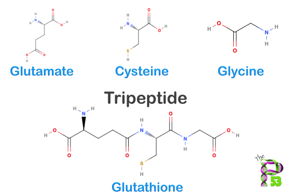
Glutathione is a tripeptide, composed of three amino acids:

  • Glutamate
  • Cysteine (the critical sulfur donor)
  • Glycine

Its uniqueness lies in the γ-glutamyl bond connecting glutamate to cysteine. This unusual bond makes glutathione resistant to most proteases, giving it stability inside cells.

The cysteine component carries a thiol (-SH) group, the chemically reactive part of the molecule. This sulfhydryl group can donate electrons, neutralizing reactive oxygen species (ROS) and other oxidants.

Glutathione exists in two major forms:

  • Reduced glutathione (GSH) – the active antioxidant
  • Oxidized glutathione (GSSG) – formed when two GSH molecules join via a disulfide bond

The GSH:GSSG ratio is a critical biomarker of oxidative stress. Healthy cells maintain a high ratio of reduced glutathione, while a shift toward oxidized glutathione signals cellular damage.

Glutathione Biosynthesis
Glutathione is synthesized inside cells through a two-step, ATP-dependent pathway:

  1. γ-Glutamylcysteine synthetase (glutamate-cysteine ligase) joins glutamate and cysteine.
    • This is the rate-limiting step.
    • Cysteine availability is the main bottleneck since dietary cysteine is less abundant.
  2. Glutathione synthetase adds glycine to complete the tripeptide.

Because ATP is required, glutathione production is tied to energy status. In times of oxidative stress, expression of γ-glutamylcysteine synthetase is boosted through the Nrf2 antioxidant response pathway, increasing glutathione synthesis.

Pathways Involving Glutathione
1. Redox Regulation and ROS Neutralization
Glutathione directly controls oxidative stress through the glutathione peroxidase (GPx) system:

  • GPx reduces hydrogen peroxide (H₂O₂) and lipid peroxides by using GSH as an electron donor.
  • GSH is oxidized to GSSG in the process.
  • Glutathione reductase then recycles GSSG back to GSH using NADPH.

This cycle prevents lipid peroxidation, protein oxidation, and DNA strand breaks. It is one of the most important antioxidant defenses in biology.

2. Detoxification and Phase II Conjugation

Glutathione plays a leading role in detoxification through glutathione S-transferase (GST) enzymes:

  • GST attaches glutathione to electrophilic toxins, drugs, and carcinogens.
  • This conjugation makes the compounds water-soluble so they can be excreted in bile or urine.
  • Heavy metals, pesticides, and pollutants are often cleared this way.

A well-known clinical example is acetaminophen overdose. When the liver runs out of glutathione to detoxify acetaminophen’s reactive metabolite (NAPQI), the toxin binds to proteins and causes severe liver damage.

3. Mitochondrial Protection

Mitochondria are both the main source of cellular energy and the main source of reactive oxygen species. Glutathione is the primary mitochondrial antioxidant:

  • It preserves mitochondrial DNA from oxidative mutations.
  • It protects the respiratory chain enzymes from thiol oxidation.
  • It maintains the mitochondrial membrane potential necessary for ATP production.

Without mitochondrial glutathione, cells are vulnerable to apoptosis or necrosis due to energy failure.

4. Immune Function and Inflammation Control

Glutathione is critical for a balanced immune system:

  • It supports T-cell and natural killer cell activity.
  • It regulates antigen presentation in immune cells.
  • It influences cytokine release, preventing both deficiency and excessive inflammation.

When glutathione levels drop, immune responses can become impaired or dysregulated, leading to either weakened defense or chronic inflammation.

5. Nitric Oxide and Vascular Function

Glutathione interacts with nitric oxide (NO) to form S-nitrosoglutathione (GSNO), which regulates NO bioavailability:

  • Helps maintain blood vessel dilation and normal blood pressure.
  • Prevents inappropriate platelet aggregation.
  • Supports endothelial cell health and vascular flexibility.

Disruption of this system contributes to hypertension and cardiovascular disease.

6. Protein Folding and Redox Signaling

In the endoplasmic reticulum (ER), glutathione contributes to protein folding by regulating disulfide bond formation. It also modifies proteins through S-glutathionylation, a reversible reaction attaching glutathione to cysteine residues on enzymes and receptors.

This modification fine-tunes signaling pathways, controlling cell proliferation, apoptosis, and stress responses.

Glutathione and Human Health
Because of its central role, glutathione depletion or imbalance is linked to many conditions:

  • Neurodegenerative disorders: Parkinson’s, Alzheimer’s, and ALS all show reduced glutathione levels in affected brain regions, leading to increased oxidative stress and neuron loss.
  • Cancer: Glutathione helps protect DNA from mutations, but cancer cells often hijack glutathione pathways to resist chemotherapy.
  • Cardiovascular disease: Low glutathione contributes to endothelial dysfunction, arterial plaque formation, and impaired nitric oxide signaling.
  • Aging: The glutathione pool declines naturally with age, weakening antioxidant defenses and increasing susceptibility to chronic illness.
  • Liver disease: Alcohol, drugs, and toxins place a heavy demand on liver glutathione. Chronic depletion contributes to fatty liver, fibrosis, and cirrhosis.

Supporting Glutathione Levels
Since cysteine is the rate-limiting factor in glutathione synthesis, dietary intake of sulfur-containing nutrients is critical. Ways to support healthy glutathione include:

  • Foods rich in sulfur compounds: garlic, onions, leeks, and cruciferous vegetables (broccoli, kale, Brussels sprouts).
  • Plant protein sources of methionine and cysteine: lentils, sunflower seeds, oats, beans.
  • Cofactors for glutathione recycling:
    • Vitamin C (recycles oxidized glutathione)
    • Vitamin E (works synergistically with GSH)
    • Selenium (essential for glutathione peroxidase function)
    • Alpha-lipoic acid (enhances glutathione regeneration)

Lifestyle choices also influence glutathione status. Chronic stress, environmental toxins, alcohol use, smoking, and poor diet all deplete glutathione reserves.

Glutathione is far more than a simple antioxidant. It is a metabolic hub that integrates redox regulation, detoxification, mitochondrial protection, immune balance, vascular health, and protein signaling. Its unique sulfur chemistry gives it unparalleled ability to protect the body against oxidative and toxic damage.

Declining glutathione is strongly associated with aging and chronic disease, while optimal levels support resilience, energy, and long-term health. Maintaining glutathione through proper nutrition, lifestyle, and metabolic support is one of the most powerful ways to safeguard cellular health.

Why Fruit Is Good for You if You Have Cancer 

By David W. Brown

The Truth About Fructose and Processed Sugar
One of the most persistent myths in cancer nutrition is the idea that “sugar feeds cancer.” This oversimplification has led some doctors and alternative health practitioners to warn patients against eating fruit altogether. Because fruit contains sugar—primarily fructose—the assumption is that it must therefore fuel tumor growth. But this view ignores decades of scientific evidence showing that whole fruits provide antioxidants, fiber, and phytonutrients that protect against cancer.

Part of the reason this myth persists is that most oncologists receive very little formal training in nutrition. A national review of U.S. medical schools found that nutrition education averages fewer than 25 hours over four years of training—less than 1% of total coursework (Adams et al., 2015). Surveys of oncologists confirm that fewer than 20% feel confident in providing nutrition advice to their patients (Kwan et al., 2018; McWhorter et al., 2022). This gap leaves many clinicians repeating simplistic phrases like “sugar feeds cancer” without the context of how whole foods, especially fruit, interact with human metabolism.

In reality, fruits are not harmful for cancer patients—they are among the most supportive foods available. They strengthen immunity, reduce inflammation, and deliver compounds that directly interfere with cancer-promoting pathways. To understand why, we need to examine how fructose in whole fruit is metabolized differently than refined sugars, and why fruit is one of nature’s most powerful allies in healing.

The Misconception: “Sugar Feeds Cancer”
All cells, both healthy and cancerous, use glucose for fuel. This has led to the popularized view that consuming sugar directly “feeds” cancer. What this overlooks is that the source and context of sugar matter enormously. Refined sugars—like high-fructose corn syrup, table sugar, and processed sweeteners—are stripped of fiber and nutrients. They rapidly spike blood glucose and insulin, creating metabolic conditions that may promote tumor growth (Johnson et al., 2007).

By contrast, whole fruits deliver natural sugars in a balanced package: water, fiber, vitamins, minerals, and bioactive compounds. This matrix slows absorption, prevents sharp glucose spikes, and provides protective substances that combat the very processes cancer depends on, such as oxidative stress and chronic inflammation (Slavin & Lloyd, 2012).

How Fructose in Fruit Is Metabolized Differently
Fructose is one of the natural sugars in fruit, alongside glucose. Processed foods often use refined fructose or high-fructose corn syrup, which behaves very differently from the fructose in whole fruit:

  1. Absorption and Fiber Modulation
    • In fruit, fructose is bound up with fiber. Fiber slows digestion and absorption, so fructose enters the bloodstream gradually, avoiding the rapid surges that processed sugars cause.
    • Processed sugars, lacking fiber, flood the liver with fructose all at once, overwhelming metabolism and contributing to fat accumulation and insulin resistance (Tappy & Lê, 2010).
  2. Liver Metabolism Pathways
    • Small amounts of fructose from fruit are easily handled by the liver, converted into glucose or stored as glycogen for later use (Mayes, 1993).
    • Large amounts of refined fructose from sodas or candies push the liver into overdrive, increasing lipogenesis (fat creation) and promoting metabolic dysfunction linked to cancer progression (Stanhope & Havel, 2010).
  3. Nutrient Synergy
    • Fruits deliver antioxidants like vitamin C, carotenoids, and polyphenols, which counteract free radicals. This protects DNA from mutations that fuel cancer growth (Lobo et al., 2010).
    • Processed sugars, by contrast, supply empty calories and can deplete the body of magnesium and B vitamins needed for cellular defense (Nielsen, 2010).

Fruit and Cancer: Protective Nutrients at Work

  1. Antioxidants and DNA Protection
    Fruits like berries, oranges, and grapes are rich in antioxidants that neutralize free radicals before they damage DNA. Blueberries, for example, contain anthocyanins that reduce oxidative DNA damage in human studies (Basu et al., 2010).
  2. Anti-Inflammatory Effects
    Chronic inflammation creates an environment where cancer can thrive. Fruits such as cherries, pineapples, and citrus contain compounds like quercetin, bromelain, and flavanones that actively lower inflammatory markers (Pan et al., 2010).
  3. Fiber and Gut Health
    Soluble and insoluble fibers in fruits not only slow sugar absorption but also nourish beneficial gut bacteria. These microbes produce short-chain fatty acids like butyrate, which have anti-cancer effects in the colon (Louis & Flint, 2017).
  4. Immune System Support
    Vitamin C from citrus boosts immune cell function, improving the body’s ability to detect and destroy malignant cells (Carr & Maggini, 2017).
  5. Detoxification Pathways
    Fruits provide phytochemicals like ellagic acid and resveratrol, which enhance the body’s detoxification of carcinogens and can directly slow tumor cell proliferation (Seeram, 2008; Bishayee et al., 2010).

Evidence from Research
Large-scale studies consistently show that higher fruit consumption is associated with reduced cancer risk and improved survival:

  • A meta-analysis in the International Journal of Cancer found that high fruit intake lowers the risk of cancers of the lung, stomach, and esophagus (Riboli & Norat, 2003).
  • The World Cancer Research Fund (2018) recommends fruit as part of cancer-preventive diets due to its fiber and phytonutrient content.
  • Specific compounds like resveratrol in grapes and ellagic acid in pomegranates have demonstrated anti-tumor activity in laboratory and animal studies (Bishayee et al., 2010).

Why Processed Sugars Are the Real Concern

While fruits protect, processed sugars harm:

  1. Insulin and IGF-1 Spikes
    Processed sugars raise insulin and insulin-like growth factor 1 (IGF-1), both of which can promote cancer cell growth and survival (Pollak, 2008).
  2. Metabolic Syndrome and Obesity
    Refined sugars drive weight gain, fatty liver disease, and systemic inflammation—all conditions that increase cancer risk and worsen outcomes (Bray & Popkin, 2014).
  3. No Protective Nutrients
    Processed sugar offers no fiber, antioxidants, or minerals to balance its effects. Instead, it depletes the body of nutrients during metabolism (Fine et al., 2012).

Practical Guidance for Cancer Patients

  1. Choose Whole Fruits – Eat fruits in their natural form rather than juices or sweetened products.
  2. Variety Matters – Aim for a rainbow of colors daily to capture diverse phytochemicals.
  3. Pair with Balanced Meals – Combine fruits with vegetables, legumes, and nuts for better absorption and satiety.
  4. Moderation, Not Elimination – There is no evidence that moderate fruit intake fuels cancer; cutting it out risks nutrient deficiencies.

The fear that “fruit sugar feeds cancer” is a misunderstanding. While refined sugars in processed foods may create metabolic conditions favorable to cancer, the fructose in fruit is metabolized differently, buffered by fiber, and delivered with a vast array of protective nutrients.

Fruits provide antioxidants, anti-inflammatory compounds, immune-boosting vitamins, and gut-friendly fiber—all of which help prevent and manage cancer. Far from being an enemy, fruit is one of nature’s most powerful allies in the fight against cancer. Patients should feel encouraged to enjoy whole fruits daily as part of a nutrient-rich, plant-based diet that supports healing and long-term wellness.

Why Aluminum Is Harmful and Serves No Purpose in the Human Body

By David W. Brown

The human body is a finely tuned biological system that depends on a wide variety of essential minerals to function properly. Iron carries oxygen through the blood. Magnesium regulates nerve signaling and muscle contraction. Zinc supports immune function and wound healing. These elements play critical roles in life and health. But aluminum is different. Despite being one of the most abundant metals in Earth’s crust and widely present in modern life—from cookware to canned foods, antiperspirants, vaccines, and processed products—aluminum serves no useful role in the human body. In fact, it is increasingly recognized as harmful, with evidence linking it to oxidative stress, inflammation, and chronic diseases.

This article explores why aluminum is not needed in human biology, how it harms the body at the cellular and systemic levels, and why reducing exposure is an important step for health.

Aluminum Has No Biological Role

Unlike calcium, potassium, and trace minerals such as selenium and manganese, aluminum is not required for any enzyme function, structural component, or biochemical pathway. The body has no transport proteins dedicated to aluminum, no storage mechanisms for beneficial use, and no receptors that recognize it as a nutrient. This alone establishes aluminum as an unnecessary and potentially disruptive substance in human physiology.

When aluminum enters the body—whether through ingestion, inhalation, or injection—it acts as a foreign metal. Instead of supporting health, it interferes with critical processes, binding to proteins and enzymes in ways that block their normal function.

Pathways of Entry into the Body

Aluminum exposure is nearly unavoidable in the modern world because of its widespread industrial and commercial use. Some of the most common pathways include:

  1. Food and Drink – Aluminum leaches from cookware, foil, and beverage cans. Processed foods, baking powders, and even some flour contain aluminum-based additives.
  2. Water Supply – Many municipal water treatment plants use aluminum salts to remove impurities, leaving residues that can be ingested daily.
  3. Personal Care Products – Most conventional antiperspirants contain aluminum salts such as aluminum chloride, aluminum chlorohydrate, or aluminum zirconium. These compounds are added because they block sweat ducts, reducing perspiration. While effective for controlling sweat, they introduce a significant source of aluminum exposure.
  4. Medical Sources – Certain vaccines contain aluminum-based adjuvants. These compounds are deliberately added to enhance immune response, making the vaccine more effective. However, they also create a direct pathway for aluminum to bypass the body’s natural barriers and enter the bloodstream. Unlike dietary aluminum, which is filtered by the gut, injected aluminum can be distributed to tissues almost immediately. In addition, some medications (such as antacids) include aluminum hydroxide.
  5. Environmental Exposure – Industrial emissions, occupational dust, and contaminated soil contribute to inhaled or ingested aluminum.

The problem is not just occasional exposure. Because the body has no efficient system for utilizing or excreting aluminum, it tends to accumulate in tissues over time, especially in the brain, bones, and kidneys.

Aluminum in Vaccines

Aluminum adjuvants in vaccines are designed to stimulate the immune system. They may have been used for decades, but their long-term effects reveal real dangers to human health. The concern is that once injected, aluminum can circulate in the blood, bind to proteins, and eventually deposit in sensitive organs.

  • Immune Activation: Aluminum particles can persist at the injection site, creating ongoing immune stimulation.
  • Systemic Distribution: Some injected aluminum binds to transferrin and albumin in the blood, carrying it to distant tissues including the brain.
  • Potential Autoimmunity: Chronic immune activation from aluminum adjuvants has been suggested as a possible trigger in certain autoimmune disorders.

Although regulatory agencies consider aluminum adjuvants “safe at current levels,” research shows that even small amounts can accumulate in tissues over years, especially when combined with aluminum from food, water, and personal care products.

Aluminum and the Brain

Perhaps the greatest concern with aluminum exposure is its effect on the nervous system. Multiple studies suggest a link between aluminum accumulation and neurodegenerative conditions such as Alzheimer’s disease, Parkinson’s disease, and amyotrophic lateral sclerosis (ALS).

  • Crossing the Blood-Brain Barrier: Aluminum can cross the blood-brain barrier by binding to transferrin, a protein normally responsible for carrying iron. Once inside the brain, it interferes with neuronal signaling.
  • Amyloid Plaques: Aluminum has been found in the amyloid plaques that characterize Alzheimer’s disease. While not the sole cause of the disease, it appears to worsen oxidative stress and inflammation in brain tissue.
  • Mitochondrial Damage: Neurons rely heavily on mitochondria for energy. Aluminum disrupts mitochondrial activity, leading to less ATP production and more free radical generation. This damages neurons and accelerates cognitive decline.

Because the brain is particularly vulnerable to toxins, aluminum buildup can have long-term consequences for memory, learning, and overall neurological health.

Aluminum and the Kidneys

The kidneys are the primary organs responsible for filtering waste and toxins from the blood. However, they are also a major site for aluminum accumulation, which places them under direct stress.

  • Kidney Retention: Healthy kidneys excrete some aluminum, but chronic exposure can overwhelm their capacity, leading to buildup.
  • Dialysis Patients at Risk: Patients with chronic kidney disease, particularly those on dialysis, are especially vulnerable. In the past, contaminated dialysis fluids caused widespread aluminum poisoning, leading to anemia, bone disease, and dementia-like symptoms.
  • Oxidative Stress: Aluminum-induced free radicals damage kidney tissue, impairing filtration and increasing the risk of chronic kidney disease progression.

Cellular and Molecular Damage from Aluminum

On a cellular level, aluminum acts as a pro-oxidant rather than an antioxidant. It promotes damage instead of preventing it. Key mechanisms include:

  1. Oxidative Stress – Aluminum increases the production of reactive oxygen species (ROS), damaging DNA, proteins, and lipids.
  2. Enzyme Disruption – Aluminum binds to critical enzymes, altering their structure and preventing normal function in energy production and detoxification.
  3. DNA Damage – By binding to phosphate groups, aluminum interferes with DNA repair mechanisms, increasing mutation rates.
  4. Immune System Dysregulation – Aluminum adjuvants overstimulate the immune system in some cases, potentially contributing to autoimmune conditions.

This cellular disruption is why aluminum toxicity can manifest in multiple organ systems, from the brain to the bones to the kidneys.

Links to Chronic Diseases

Research increasingly connects aluminum exposure to a variety of chronic illnesses:

  • Alzheimer’s Disease: Elevated aluminum levels have been measured in the brains of Alzheimer’s patients.
  • Parkinson’s Disease: Aluminum may exacerbate dopaminergic neuron loss, a hallmark of Parkinson’s.
  • Cancer: Although research is ongoing, some studies suggest aluminum compounds may play a role in breast cancer when absorbed through antiperspirants.
  • Autoimmune Disorders: Excessive immune stimulation from aluminum adjuvants is under investigation for potential links to autoimmune conditions.

While not always the sole cause, aluminum often acts as a co-factor that worsens disease risk and progression.

Why the Body Cannot Use Aluminum

The clearest evidence that aluminum is harmful lies in the fact that the human body has no biological pathways that require it. Essential minerals have dedicated roles—iron carries oxygen, magnesium activates over 300 enzymes, zinc regulates gene expression. Aluminum has none.

Instead of supporting life, it mimics or replaces beneficial metals in harmful ways. For instance, by binding to iron-binding proteins, it disrupts iron transport. By interfering with calcium signaling, it weakens bones. By damaging mitochondria, it robs cells of energy.

Thus, aluminum is not just useless—it is actively disruptive.

Reducing Aluminum Exposure

While complete avoidance is nearly impossible, several steps can help minimize exposure:

  1. Cookware Choices: Use stainless steel, glass, or cast iron instead of aluminum pans or foil.
  2. Food Labels: Avoid processed foods containing aluminum-based additives such as sodium aluminum phosphate.
  3. Water Filters: Invest in filtration systems that reduce aluminum levels in tap water.
  4. Personal Care: Choose aluminum-free deodorants and natural body care products.
  5. Medical Awareness: Discuss aluminum exposure with healthcare providers, especially if using antacids or undergoing dialysis.

These practical steps can significantly lower the toxic burden on the body.

Aluminum is one of the most abundant metals in the environment, but it has no rightful place in the human body. Unlike essential minerals, it plays no beneficial role in biology. Instead, it disrupts cellular function, promotes oxidative stress, and accumulates in vulnerable organs such as the brain, bones, and kidneys. Over time, this accumulation contributes to neurodegeneration, skeletal weakness, kidney disease, and potentially cancer and autoimmune disorders.

The human body does not need aluminum—ever. Its presence is only harmful, and its effects become more dangerous with chronic exposure. Recognizing aluminum for what it is—a toxic, non-essential metal—empowers us to take steps to minimize contact and protect long-term health.

Nearly 40% of Autistic Children Deficient in Vitamin D and Iron

By David W. Brown

A growing body of research has documented that children on the autism spectrum are at higher risk for select micronutrient shortfalls—especially vitamin D and iron—owing to sensory-based food selectivity, limited dietary variety, and less outdoor time (which lowers skin production of vitamin D). The new Singapore cohort adds careful numbers to that picture: out of 222 autistic children who had vitamin D measured, 36.5% were insufficient/deficient; out of 236 who had iron studies, 37.7% had iron deficiency; and 15.6% of those with full blood counts had iron-deficiency anemia. Importantly, “picky eating” did not reliably predict who was deficient—meaning clinicians shouldn’t wait for pronounced feeding selectivity to screen.

These findings echo prior evidence. Earlier work has linked autism with lower vitamin D status and, in many cohorts, higher rates of iron deficiency compared with neurotypical controls. Recent reviews suggest that vitamin D status can correlate with symptom severity and that correcting deficiency may improve certain outcomes, though larger, longer trials are still needed. 

Why vitamin D and iron matter

Vitamin D supports calcium–phosphate balance, bone health, immune regulation, and neurodevelopment. Food sources are limited; beyond sunlight, typical contributors are mushrooms (especially UV-exposed). Many public-health bodies note that routine vitamin D supplementation is often appropriate for children and other groups given widespread insufficiency, particularly when sunlight exposure is low. 

Iron is essential for oxygen transport and for enzymes that shape attention, learning, memory, and motor development. Plant foods provide nonheme iron (lentils, beans, tofu/tempeh, pumpkin seeds, cashews, quinoa, dark leafy greens). Pairing plant iron with vitamin-C–rich foods (citrus, berries, peppers, tomatoes, broccoli) significantly boosts uptake. 

Where a Whole-Food, Plant-Forward Pattern Fits—Including the P53 Diet


A thoughtfully planned whole-food, plant-forward diet—like the P53 Diet framework—emphasizes fruits, vegetables, legumes, whole grains, nuts, and seeds while removing ultra-processed items. Two big advantages of this pattern for families supporting autistic children:

  1. Higher nutrient density and fiber for the calories consumed. Diverse plant foods deliver broad micronutrient coverage (folate, magnesium, potassium, many phytonutrients) that typical “beige” kid diets lack. Large analyses show that shifting intake toward plant foods and away from red/processed meats is associated with better cardiometabolic profiles and lower risk of chronic disease over time—benefits that matter for the whole household. 
  2. Built-in opportunities to optimize iron and vitamin D—if you’re intentional.
    • You can reach iron needs with legumes (lentils, chickpeas, black beans), soy foods, seeds (pumpkin, sesame), nuts, dark greens, and fortified whole grains—and by routinely pairing them with vitamin-C-rich produce to magnify absorption.
    • Vitamin D remains the exception: sunlight plus plant milks, often still won’t meet needs year-round; a supplement is commonly recommended for children by several expert groups. Your plan should treat vitamin D like a “must-check” nutrient. GET YOUR KIDS OUTSIDE MORE TO GET THEIR VITAMIN D!

Practical P53-style strategies for families

  • Make the plate colorful and predictable. Sensory preferences are real. Offer a reliable structure (same plate/bowl, consistent mealtime cues) but vary the colors and textures within that structure—e.g., red lentil pasta with tomato-pepper sauce one night; chickpea pasta with lemon-broccoli another. Consistency lowers mealtime stress while nudging variety.
  • Load iron + vitamin C together. Chili with black beans (iron) + diced tomatoes/bell peppers (vitamin C). Hummus (iron) + citrus segments. Tofu stir-fry with broccoli and pineapple.
  • Lean on UV-exposed mushrooms. Sautéed or blended into sauces, they can add meaningful vitamin D—helpful, though still usually not enough alone. 
  • Mind the other “usual suspects.” Any plant-exclusive plan should also ensure vitamin B12 and iodine (iodized salt or nutritional yeast), with attention to calcium, zinc, and selenium as needed. 

Bringing it together: why the P53 Diet is a strong fit

The P53 Diet’s core principles—whole, minimally processed plants; high diversity; avoidance of refined oils and ultra-processed foods; and a science-first approach—map cleanly onto what the evidence suggests for optimizing everyday health while guarding against common shortfalls:

  • It raises overall diet quality, which supports healthy growth, GI function, and long-term cardiometabolic health for kids and adults alike. 
  • It makes iron coverage practical via legumes, soy, greens, seeds, and fortified grains—especially when recipes routinely pair these with vitamin-C-rich produce. 
  • It encourages a systems view: not just “fixing a number,” but improving sleep, movement, and whole-household food patterns that make nutrient sufficiency and metabolic health sustainable.

The new Nutrients study sharpens an increasingly consistent message: among children with autism who are tested, roughly four in ten can be low in vitamin D or iron, and you can’t reliably spot those kids by feeding behavior alone. As the authors note, “a significant proportion of almost 40% of children diagnosed with ASD … had vitamin D insufficiency/deficiency and/or iron deficiency.” That’s a call for routine screening and targeted nutrition support, not alarm. 

A well-planned, whole-food, plant-forward pattern—like the P53 Diet—offers a powerful foundation for families: it elevates diet quality, improves long-term health markers, and, with a few smart habits (vitamin C with plant iron), closes the exact gaps highlighted by this research. Work with your pediatrician. With an evidence-aligned approach, plant-based eating becomes not just compatible with autism-informed nutrition—but one of the most practical ways to promote overall health for your child and your household. GET YOUR KIDS OUTDOORS MORE!

Reference:
Primary study: Koh MY, Lee AJW, Wong HC, Aishworiya R. Occurrence and Correlates of Vitamin D and Iron Deficiency in Children with Autism Spectrum Disorder. Nutrients. 2025;17(17):2738. 

Why Cooking Oils Are Harmful to the Human Body

By David W. Brown

Cooking oils are marketed as everyday essentials, but scientific evidence shows they can be detrimental to human health. The issues arise from the way oils are produced, their biochemical effects in the body, and the toxic solvents used during extraction, particularly hexane.

Industrial Processing and Hexane Extraction

Most commercial cooking oils—soybean, corn, canola, sunflower, safflower, and others—are not simply “pressed” from plants. Instead, they undergo industrial solvent extraction, where the seeds are crushed and treated with hexane, a petroleum-derived chemical. Hexane is favored because it efficiently strips nearly all oil from plant material, maximizing yield. After extraction, the oil is heated to evaporate most of the hexane, but residues can remain. Even trace levels of hexane are concerning: it is classified as a neurotoxin and inhalation exposure is linked to nerve damage in workers. While regulators argue the amounts left in oil are small, chronic dietary exposure has not been thoroughly studied. Thus, oils made with hexane introduce a potential chemical contaminant into the human food supply.

Refining, Bleaching, and Deodorizing

After extraction, oils are refined, which involves neutralizing free fatty acids with lye, bleaching with clays to remove pigments, and deodorizing at very high heat to strip unpleasant odors. This high-heat treatment alters the chemical structure of fatty acids, generating trans fats and other oxidative byproducts even before the oil reaches consumers. Many of these compounds are pro-inflammatory and cytotoxic, setting the stage for long-term health consequences.

Oxidation and Free Radical Damage

Once extracted, refined oils are chemically unstable. Polyunsaturated fatty acids (PUFAs) in oils like soybean and corn are highly prone to oxidation, especially when exposed to light, air, and heat during cooking. Oxidized oils form lipid peroxides and aldehydes, which can damage DNA, proteins, and cell membranes. These compounds trigger oxidative stress and inflammation, both fundamental drivers of chronic diseases including cancer, atherosclerosis, and neurodegenerative disorders.

Distortion of Omega-6 to Omega-3 Ratio

Vegetable oils are extremely high in omega-6 fatty acids (linoleic acid) but nearly devoid of omega-3s. While omega-6 fats are essential in small amounts, modern diets laden with cooking oils push the ratio of omega-6 to omega-3 far beyond the ideal balance (often 20:1 versus the recommended 1–4:1). Excess omega-6 promotes the production of pro-inflammatory molecules called eicosanoids, fueling systemic inflammation that underlies arthritis, cardiovascular disease, diabetes, and obesity.

Impact on Human Metabolism

Refined oils are calorie-dense but nutrient-poor, offering no fiber, vitamins, or minerals. They represent “empty calories” that disrupt satiety signals and contribute to weight gain. Furthermore, heating oils during frying produces advanced lipid oxidation end products (ALEs), which impair insulin signaling and promote insulin resistance. This helps explain the strong association between frequent fried food consumption and type 2 diabetes.

Cooking oils may seem harmless, but their risks are embedded at every stage: toxic solvent extraction with hexane, chemical refining and deodorizing, oxidative instability, omega-6 overload, and pro-inflammatory byproducts formed during cooking. Regular consumption exposes the human body to free radical damage, chronic inflammation, and toxic residues, which together contribute to obesity, diabetes, cardiovascular disease, and cancer. For optimal health, whole plant foods such as nuts, seeds, and avocados provide natural fats along with fiber, vitamins, and antioxidants—delivering the benefits of healthy fats without the hazards introduced by industrially processed oils.

The Health Risks of the Carnivore Diet and the Superiority of a Plant-Based Diet

By David W. Brown

The carnivore diet—a dietary regimen consisting entirely of animal-based foods, typically red meat, organ meats, and animal fats—has gained popularity among proponents seeking weight loss, simplicity, or relief from autoimmune conditions. Advocates often claim that this zero-carb, high-protein lifestyle mimics the dietary habits of early humans and provides a powerful antidote to modern metabolic disorders. However, mounting scientific evidence paints a very different picture. The carnivore diet, though potentially beneficial in the short term for specific conditions, carries significant long-term health risks. These risks span cardiovascular, renal, gastrointestinal, hormonal, and oncological domains.

In contrast, a well-balanced plant-based diet—especially one rich in whole foods such as fruits, vegetables, legumes, whole grains, seeds, and nuts—has repeatedly demonstrated its power in preventing, managing, and even reversing chronic diseases. This article details the health issues associated with a carnivore diet, explains the underlying biological pathways, and outlines why a plant-based diet remains the healthiest and most sustainable nutritional approach.

Cardiovascular Risks of the Carnivore Diet
Elevated LDL Cholesterol and Atherosclerosis
The carnivore diet is rich in saturated fats and cholesterol. Consumption of these nutrients leads to an increase in low-density lipoprotein (LDL) cholesterol, the “bad” cholesterol, which is directly implicated in the development of atherosclerosis. Atherogenesis begins with damage to the endothelial lining of blood vessels. LDL particles penetrate the endothelium and become oxidized (oxLDL), triggering an immune response that recruits macrophages. These immune cells engulf the oxLDL, becoming foam cells and forming fatty streaks, which are the precursor to plaques that narrow arteries and reduce blood flow.

A meta-analysis of 395 prospective studies found that high LDL is causally related to atherosclerosis and coronary artery disease (Ference et al., 2017). Diets high in red and processed meat also correlate with a greater risk of cardiovascular mortality (Micha et al., 2012).

Impaired Nitric Oxide Synthesis
Endothelial function depends on nitric oxide (NO), a molecule that relaxes blood vessels. Animal proteins lack nitrates, which are abundant in green leafy vegetables and are precursors to nitric oxide. A carnivore diet reduces NO synthesis, leading to vasoconstriction, hypertension, and endothelial dysfunction—key steps in cardiovascular disease.

Renal Dysfunction and Protein Overload
Glomerular Hyperfiltration
The high-protein load from a carnivore diet imposes metabolic stress on the kidneys. Increased protein intake leads to glomerular hyperfiltration—a temporary rise in kidney filtration rate that compensates for the extra nitrogen load from protein breakdown. Over time, this adaptation becomes pathological, contributing to glomerulosclerosis (scarring of glomeruli) and progressive kidney disease.

Protein metabolism produces nitrogenous wastes like urea and ammonia, which the kidneys must excrete. This increased workload accelerates renal decline in susceptible individuals, especially those with pre-existing kidney issues.

A long-term study by Knight et al. (2003) found that women with mild renal insufficiency who consumed high protein diets experienced accelerated kidney function loss.

Gastrointestinal Dysbiosis and Constipation
Lack of Fiber and Microbiome Imbalance
The carnivore diet contains no dietary fiber, an essential nutrient for feeding beneficial gut bacteria. A fiber-deficient diet leads to dysbiosis—an imbalance between good and harmful microbes. This impairs the gut barrier, promoting systemic inflammation through endotoxemia (leakage of lipopolysaccharides into the bloodstream).

Studies have shown that fiber promotes the production of short-chain fatty acids (SCFAs) like butyrate, which nourish colonocytes, reduce inflammation, and regulate immune responses. A carnivore diet inhibits SCFA production, weakening gut integrity.

Constipation is also a frequent issue due to the absence of insoluble fiber, which adds bulk to stool and facilitates intestinal motility.

Cancer Risk and Heme Iron Toxicity
Carcinogenic Compounds in Meat
Cooking meat at high temperatures generates carcinogenic heterocyclic amines (HCAs) and polycyclic aromatic hydrocarbons (PAHs). Moreover, processed meats contain nitrates and nitrites, which can form nitrosamines—a class of potent carcinogens.

The World Health Organization (2015) classified processed meat as a Group 1 carcinogen (definitively carcinogenic to humans) and red meat as a Group 2A carcinogen (probably carcinogenic). Increased consumption is strongly associated with colorectal, pancreatic, and prostate cancers.

Heme Iron and Oxidative Stress
Heme iron from animal sources catalyzes the formation of reactive oxygen species (ROS), which damage DNA and promote carcinogenesis. Unlike non-heme iron from plants, heme iron bypasses homeostatic controls, leading to iron overload and oxidative stress.

Hormonal and Endocrine Disruption
Insulin Resistance and IGF-1
A carnivore diet, while often low in carbohydrates, promotes excess amino acid intake that stimulates insulin and insulin-like growth factor 1 (IGF-1). IGF-1 promotes cell proliferation and inhibits apoptosis—contributing to cancer risk.

High levels of IGF-1 have been linked to increased risk of breast, prostate, and colorectal cancers. Additionally, diets low in carbohydrates but high in fat can paradoxically worsen insulin sensitivity over time due to ectopic fat accumulation in muscle and liver cells.

Thyroid Suppression
Low carbohydrate intake on the carnivore diet can reduce triiodothyronine (T3) levels, leading to symptoms of hypothyroidism including fatigue, cold intolerance, and hair thinning. This occurs because carbohydrates are required to convert thyroxine (T4) into its active form, T3.

Nutrient Deficiencies
Despite claims that organ meats supply all necessary nutrients, the carnivore diet lacks many critical micronutrients:

  • Vitamin C: Essential for collagen synthesis and immune function. Absence leads to scurvy, fatigue, and poor wound healing.
  • Magnesium: Critical for over 300 enzymatic reactions. Deficiency can lead to muscle cramps, arrhythmias, and depression.
  • Folate: Vital for DNA synthesis and red blood cell formation. Deficiency causes anemia and neural tube defects in pregnancy.
  • Fiber: Essential for bowel health and glycemic control.
  • Phytochemicals: Plant-based compounds like flavonoids and carotenoids have antioxidant, anti-inflammatory, and anti-cancer effects.

Chronic Inflammation and Autoimmunity
While some individuals report symptom relief from autoimmune diseases on a carnivore diet, this is often a result of eliminating processed foods and allergens, not due to meat consumption itself. Over time, the lack of anti-inflammatory plant compounds may increase systemic inflammation.

High intake of red meat elevates levels of TMAO (trimethylamine N-oxide), a metabolite linked to atherosclerosis and inflammation. TMAO is formed by gut bacteria when digesting carnitine and choline—abundant in red meat.

Plant-Based Diet
The Case for a Whole-Food, Plant-Based Diet

Cardiovascular Health
A plant-based diet has consistently been shown to reverse heart disease, reduce blood pressure, and lower LDL cholesterol. Dr. Caldwell Esselstyn and Dr. Dean Ornish demonstrated that a low-fat, plant-based diet, combined with lifestyle changes, can halt and reverse coronary artery disease.

Leafy greens, legumes, fruits, and nuts contain natural nitrates, antioxidants, and polyphenols that support endothelial function and reduce oxidative stress.

Cancer Prevention
The World Cancer Research Fund and American Institute for Cancer Research recommend a diet high in plant foods to reduce cancer risk. Cruciferous vegetables (e.g., broccoli, kale) contain sulforaphane, which induces detoxification enzymes and suppresses tumor growth. Flaxseeds provide lignans that lower breast cancer risk by modulating estrogen metabolism.

Gut Health and Immunity
Plant foods nourish a diverse microbiome. Fiber-rich diets increase SCFA production, reduce gut inflammation, and improve mucosal immunity. Fermented plant foods (e.g., kimchi, sauerkraut) also enhance microbiota diversity and gut resilience.

Hormonal Balance
Plant-based diets naturally regulate insulin and IGF-1 levels. Lower fat intake improves insulin sensitivity, and whole grains provide steady glucose release without spikes.

Soy foods, often demonized, are actually protective—containing isoflavones that modulate estrogen receptors and reduce cancer risk, particularly in postmenopausal women.

Anti-inflammatory and Antioxidant Effects
Plants are rich in vitamins C and E, beta-carotene, quercetin, and resveratrol—compounds that neutralize free radicals and reduce systemic inflammation. These effects have been shown to reduce arthritis symptoms, improve brain function, and support longevity.

Sustainability and Ethical Considerations
A plant-based diet is not only healthier but also more sustainable. Livestock farming is a leading contributor to greenhouse gases, deforestation, and water pollution. Transitioning to plant-based eating reduces environmental impact and aligns with ethical treatment of animals.

The carnivore diet may provide short-term symptom relief for some individuals, particularly those with severe food sensitivities. However, it is inherently deficient in fiber, phytochemicals, and numerous vitamins. It promotes cardiovascular disease, cancer, and renal stress through mechanisms involving LDL cholesterol, oxidative damage, hormone dysregulation, and gut microbiome disruption.

In contrast, a whole-food, plant-based diet nourishes every system in the human body, providing comprehensive protection against modern chronic diseases. It supports cardiovascular function, regulates hormones, fosters a healthy gut, boosts immunity, and prevents cancer. Moreover, it offers a sustainable and ethical approach to nutrition that aligns human health with planetary well-being.

The best path forward for long-term health is to embrace a colorful, diverse, and fiber-rich plant-based diet that fuels both vitality and longevity.